This volume covers all aspects of bone scintigraphy. It traces radiopharmaceutical development, scanning technique, and interpretation. It includes sections on bone marrow imaging and even on DEXA imaging for bone densitometry (since this technique began as dual photon absorptiometry (DPA), and has remained in nuclear medicine at many institutions).
There are chapters on the many uses of the bone scan: Tumor imaging (metastatic disease and primary bone tumor imaging and differential diagnosis), infection imaging, orthopedic complications, sports injuries, and a number of other fairly unique issues. There is a chapter on “conditions that result in a normal bone scan” and “conditions that are cold on bone scan”, which are unique important differential diagnoses in skeletal scintigraphy that all readers should be well versed in. There is a very important section on “soft tissue uptake on bone scan”, which is important clinically, but also, very frequent fodder for board exams.
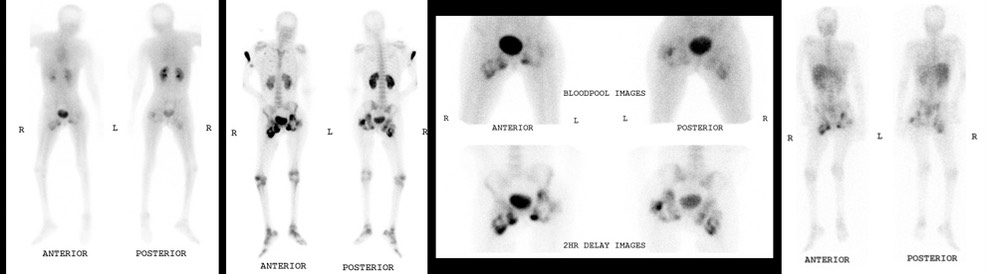
There are lots of images, particularly of the “aunt minnies” in skeletal scintigraphy that might show up on the core exam.
Table of Contents:

Representative Images, from this image rich volume:
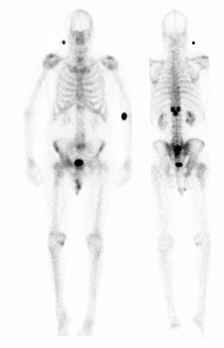
Classic gloved hand (actually just 2 fingers, radial artery injection) of an intra-arterial injection.
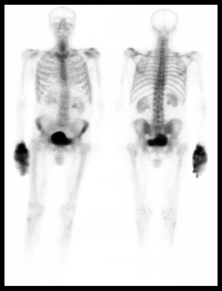
The “Mickey Mouse” sign (highly appropriate from a Florida based practice: “I’m going to DisneyWorld”). Also shows Paget’s of the sacrum.
The bone scan pattern of calciphylaxis (and also the pattern of nephrogenic system fibrosis (NSF):
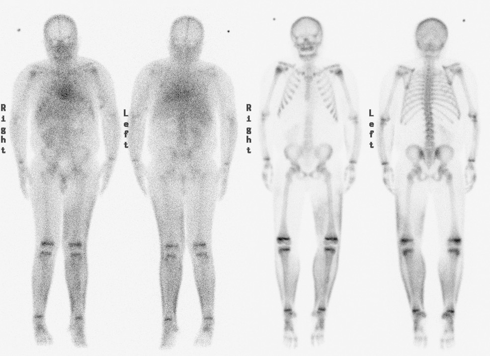
The bone scan appearance of “Myositis Ossificans of Paraplegia”.