My early career was academically as a GI nuclear medicine physician, with an academic focus on GI motility (the esophagus, the stomach, the small bowel). All-the-while, my clinical practice is heavily GI oriented. (I grew up when GI fluoroscopy was “king”, involved with a site that performed hundreds of GI exams every single day, in the days of aggressive double-contrast UGI and barium enema exams, as well as enteroclysis. I established CT enterography, MR enterography, Virtual Colonography, and MR defecography at my current institution and continue to be active in this arena). This volume reflects this broad experience, and discuss the common and the very unusual exams that nuclear medicine can be involved with. It starts with the top of the GI tract, with even a review of salivary imaging, the saliva scan in infants, esophageal scintigraphy, gastric emptying studies, and even the more infrequent and unusual small bowel and colonic motility studies. I have had one of the busiest motility practices in the country, perform 10 – 20 gastric studies per day for most of my career. Not a fan of the national standard for gastric emptying study, I review the pros and cons of this exam, and cover the more atypical exams, like motility studies after bariatric surgery.
Scans for ectopic gastric mucosa (Meckel’s scans) and GI bleeding studies are covered, with a large number of bleeding study illustrations.
Liver imaging (even with a brief review of the ancient liver spleen scan) is covered, with review of more modern uses, include imaging required to monitor 90Y therapy. Biliary imaging is fully reviewed, including more unique uses, like quantitation of enterogastric reflux or using these agents to classify primary hepatic tumors. Splenic imaging with sulfur colloid or with heat-damaged RBCs is covered as well.
Overall, this is a unique prospective of the common and uncommon indications for GI scintigraphy.
REPRESENTATIVE IMAGES:
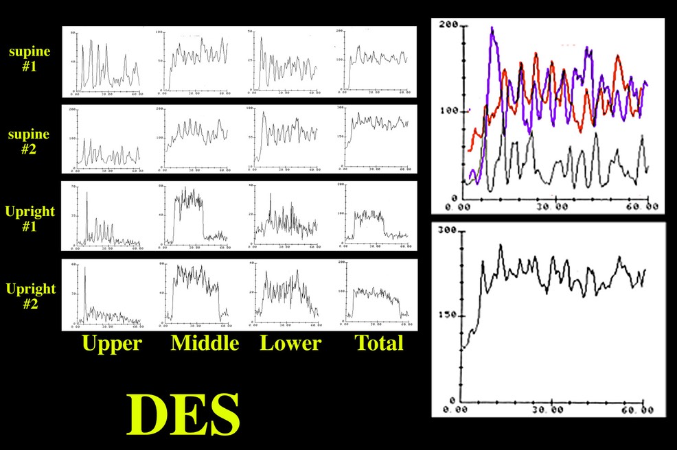
DIFFUSE ESOPHAGEAL SPASM on Esophageal Scintigraphy:
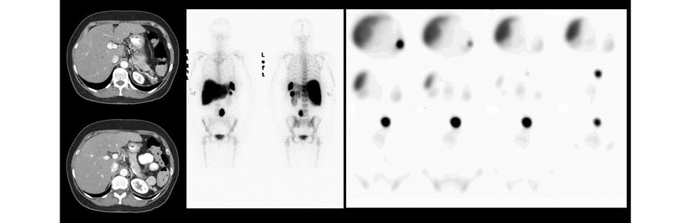
SPLENOSIS ON HEAT-DAMAGED RBC SCAN:
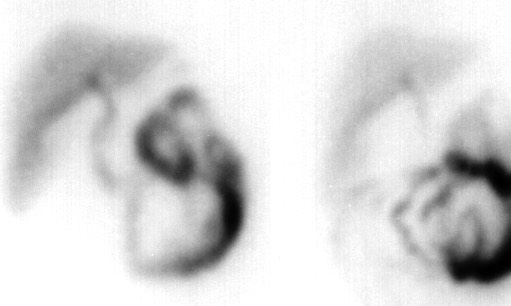
RIM SIGN of ACUTE CHOLECYSTITIS on HEPATOBILIARY SCAN:
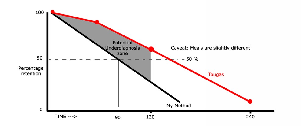
DRANE Quantitation method vs. the “National Standard”
My prettiest scan ever: Peliosis Hepatis from anabolic steroid therapy on a liver-spleen scan
ACTIVE GI BLEED ON TAGGED RBC STUDY
This browser cannot play the embedded video file.